Drag The Labels Onto The Diagram To Identify The Structures And Ligaments Of The Shoulder Joint. : Nasa Human Vestibular System In Space / Drag the labels onto the diagram to identify the muscles that move the forearm and hand, anterior view.
Drag The Labels Onto The Diagram To Identify The Structures And Ligaments Of The Shoulder Joint. : Nasa Human Vestibular System In Space / Drag the labels onto the diagram to identify the muscles that move the forearm and hand, anterior view.. Drag the appropriate labels to their respective targets. You just studied 368 terms! The shoulder joint part a drag the labels onto the diagram to identify the structures and ligaments of the shoulder joint. Just remember the articulating surfaces. Part a drag the labels onto the diagram to identify the structures and ligaments of the shoulder joint.
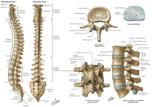
Drag the appropriate labels to their respective targets. It is the most common type of joint found in the human body, and contains several structures which are not seen in fibrous or cartilaginous joints. The anatomy of our musculoskeletal system is quite complex. Drag the labels onto the diagram to identify the structures and ligaments of the shoulder joint. Vertebral anatomy (a lateral and slightly inferior view of a vertebra) identify the structures on a vertebra.
It consists of a large number of tendons, ligaments, bones, cartilage, joints, and bursae.
Label the curves and regions of the vertebral column. Drag the appropriate labels to their respective targets. Drag the labels onto the diagram to identify the muscles that move the forearm and hand, anterior view. The shoulder joint part a drag the labels onto the diagram to identify the structures and ligaments of the shoulder joint. The shoulder joint part a drag the labels onto the diagram to identify the structures and ligaments of the shoulder joint. These ligaments stabilize the acromioclavicular (ac) joint. Shoulder ligaments also form the joint capsule that surround the glenohumeral joint. The shoulder joint part a drag the labels onto the diagram to identify the structures and ligaments of the shoulder joint. It is the most common type of joint found in the human body, and contains several structures which are not seen in fibrous or cartilaginous joints. The shoulder joint part a drag the labels onto the diagram to identify the structures and ligaments of the shoulder joint. Look at pic which joint is considered the most flexible joint in the body? These are the main ligaments that help to stabilize the joints of the shoulder: The shoulder joint part a drag the labels onto the diagram to identify the structures and ligaments of the shoulder joint.
At these joints, the rounded head of one bone (the ball) fits into the concave articulation (the socket) of the adjacent bone (see figure 9.4.3f). It consists of a large number of tendons, ligaments, bones, cartilage, joints, and bursae. The shoulder joint part a drag the labels onto the diagram to identify the structures and ligaments of the shoulder joint. Dense regular connective tissue from a tendon (500x). Drag the labels onto the diagram to identify the parts of the placenta and associated structures.

Shoulder pain and apprehension are indicative of shoulder impingement.
Part a this movement is known as __________. Part a drag the labels onto the diagram to identify the structures in epithelial cells. Identify the saddle joint of the skeleton. Shoulder ligaments also form the joint capsule that surround the glenohumeral joint. Which of the following is true regarding the structure indicated by the arrow in the joint depicted in a? We are able to control our muscles by sending stimulating impulses via nerves from our brain. Carpometacarpal joint of the thumb. Drag the appropriate labels to their respective targets. be prepared to identify all labeled skull structures in this image on upcoming exams nice work! Reset help medullary cavity spongy bone per. 8 name the arteries and the nerves that coracohumeral ligament : The shoulder joint part a drag the labels onto the diagram to identify the structures and ligaments of the shoulder joint. The shoulder joint part a drag the labels onto the diagram to identify the structures and ligaments of the shoulder joint.
Shoulder joint ligament tendon collagen fibers nuclei of fibroblasts structure of collogen each collagen fiber consists of aggregates of tropocollagen molecules. This is an online quiz called shoulder ligaments. This structure allows rotational movement, as the rounded bone moves around its own axis. Drag the appropriate labels to their respective targets. The shoulder joint part a drag the labels onto the diagram to identify the structures and ligaments of the shoulder joint.

Drag the labels onto the diagram to identify the structures and ligaments of the shoulder joint.
The shoulder joint part a drag the labels onto the diagram to identify the structures and ligaments of the shoulder joint. 8 name the arteries and the nerves that coracohumeral ligament : These are the main ligaments that help to stabilize the joints of the shoulder: Look at pic which joint is considered the most flexible joint in the body? Part a drag the labels onto the diagram to identify the curves and regions of the vertebral column. All hinge joints also contain muscles, ligaments, and other tissues that stabilize the joint. An example of a pivot joint is the joint of the first and second vertebrae of the neck that allows the head to move back and forth (figure 4). Synovial joints are characterized by the presence of a joint cavity. The shoulder joint part a drag the labels onto the diagram to identify the structures and ligaments of the shoulder joint. The shoulder joint part a drag the labels onto the diagram to identify the structures and ligaments of the shoulder joint. Identify the saddle joint of the skeleton. The glenoid fossa forms a very shallow socket, so the muscles, ligaments, and cartilage of the shoulder joint reinforce its structure and help to prevent dislocations. The joint of the wrist that allows the palm of the hand to be turned up and down is also a pivot joint.


Komentar
Posting Komentar